
Dystrophy is an inherited condition that usually develops in both eyes. Depending on the dystrophy, it can occur early in life or much later. Usually, dystrophies are slowly progressive; some decrease vision and some do not. Dystrophies can affect the surface of the cornea, the middle layers or the deepest parts. There are many types of dystrophies but the most common are:
Basement Membrane Dystrophy (also known as map-dot dystrophy or geographic dystrophy). This is the most common dystrophy of the cornea. It may be incidental (causing no problems with pain or blurred vision) or can give decreased vision or recurrent erosion syndrome.
The surface of the cornea has numerous fingerprint lines, or dots or whitish cysts. This can appear at any age but is more common over the age of 50. Usually, it is bilateral. This condition may cause an irregularity to the surface of the cornea, giving blurred or ghost-like vision.
A variety of treatment options include:
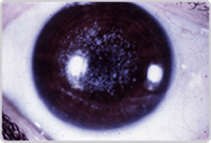
This is dominantly inherited with “granular” looking spots (Fig 1) in the cornea giving a decrease in vision or recurrent erosion syndrome. The granules are made up of a substance called hyaline.
When vision is significantly decreased, a corneal transplant is indicated.
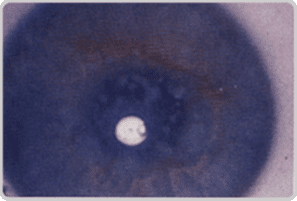
In this dominantly inherited condition, glass-like branching lines made of amyloid accumulate (Fig 2).
One can have recurrent erosion syndrome or need a corneal graft to increase vision.
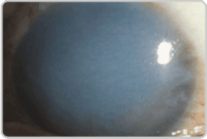
This is the least common of the middle layer dystrophies and is recessively inherited (Fig 3). It can start at as young an age as 3–9 years. A mucopolysaccharide deposits in the cornea, appearing as large white spots that can grow together causing the whole cornea to become hazier.
Treatment is similar to the above dystrophies.

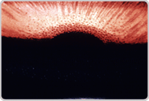
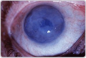
Very common, FCD is dominantly inherited and more common in females, usually starting after 50 years of age (Fig 4). There is a dysfunction in the deepest layer of the cornea when the endothelial cells die off. These are non-reproducing cells that determine how much water gets into the cornea. This affected area looks like the surface of an orange (Fig 5). As more water comes into the cornea, it makes it hazier (edema) which decreases vision.
The early symptoms are blurred vision in the morning after the eyes have been closed all night. As time goes on, more swelling comes with worse vision. At advanced levels, one can even develop “blisters” on the surface, giving a foreign body sensation and pain (Fig 6).
This condition can be treated in its early stages with salt drops or ointment that draws the water out of the cornea. Steroids can be used to help the “sick cells” work better. When severe, a full PKP or split thickness corneal transplant can be performed.
Descemet’s stripping endothelial keratoplasty, also known as DSEK, is a partial-thickness corneal transplant. Occasionally diseases, such as Fuchs’ Dystrophy, or previous eye surgery cause the loss of a thin layer of cells, called endothelial cells, on the back surface of the cornea.
In DSEK, the thin layer of damaged or unhealthy endothelial cells is removed and replaced with donor corneal tissue containing healthy endothelial cells. In contrast to full-thickness corneal transplantation, no sutures are used to secure the partial-thickness corneal transplant into place. Instead, a large air bubble is placed inside the eye, behind the transplanted tissue, which pushes the donor tissue into place.
Wheaton Eye Clinic’s unparalleled commitment to excellence is evident in our continued growth. Today we provide world-class medical and surgical care to patients in six suburban locations—Wheaton, Naperville, Hinsdale, Plainfield, St. Charles, and Bartlett.
(630) 668-8250 (800) 637-1054