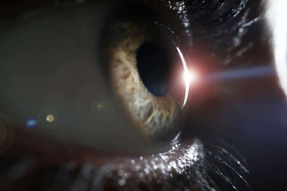
A Retinal detachment is a separation of the retina, the light sensitive layer in the back of the eye, from the eye wall. The retina functions like the film in a camera and needs to be firmly attached to the eye wall in order to see properly.
When the retina detaches, patients will perceive a curtain or shadow blocking part or all of the vision in the affected eye. Other symptoms of retinal detachment can include flashes of light or floaters in the field of view. A retinal detachment is an eye emergency and can lead to blindness if not corrected.
Retinal detachments usually develop when a tear or hole develops in the retina. The hole allows fluid to seep under the retina and lift the retina off the eye wall. Other types of retinal detachments caused by scarring or inflammation can occur, but these are much less common. Contrary to popular belief, most retinal detachments are spontaneous and unrelated to injury to the eye.
Patients with a family history of retinal problems, patients with abnormal thinning of the retina called lattice degeneration, patients who previously have had cataract surgery, or very nearsighted individuals are all at high risk for developing retinal detachment. This condition occurs in approximately 1 per 10,000 patients.
Because the natural history of retinal detachments almost always leads to blindness, the vast majority of retinal detachments are treated. Treatment always involves surgery. Several main types of treatment strategies are commonly used: pneumatic retinopexy, scleral buckle, vitrectomy or a combination approach.
This treatment uses a gas bubble to push the retina on to the back of the eye, and is only useful in a small number of patients with retinal detachment. However, it is the least invasive treatment available.
This procedure involves wrapping a plastic band around the eye to indent the eye wall to reattach the retina. Scleral buckling is a very common method of reattaching the retina and has been used for decades with excellent success rate.
This procedure involves removal of the jelly in the inside of the eye called the vitreous. Removal of the vitreous allows the retina to be reattached on the inside of the eye and has become a much more common method of reattaching both simple and complex retinal detachments in recent years.
In many cases, a combination of two or more of the above treatments are utilized together to give the greatest success in reattaching retinas.
Surgical procedures are usually carried out in the operating room under local anesthesia as an out-patient. The patient’s eye is thoroughly numbed, and the patient rests comfortably during the 30 to 90 minute procedure. Success rate for reattaching a retina following one surgical procedure is approximately 90%, and over 95% of all retinal detachments can ultimately be reattached successfully.
Wheaton Eye Clinic surgeons have extensive experience diagnosing and treating hundreds retinal detachments each year using all of the techniques listed above. Your surgeon will discuss with you which surgical procedure is best in your circumstances.
Wheaton Eye Clinic’s unparalleled commitment to excellence is evident in our continued growth. Today we provide world-class medical and surgical care to patients in six suburban locations—Wheaton, Naperville, Hinsdale, Naper Plainfield, St. Charles, and Bartlett.
(630) 668-8250 (800) 637-1054